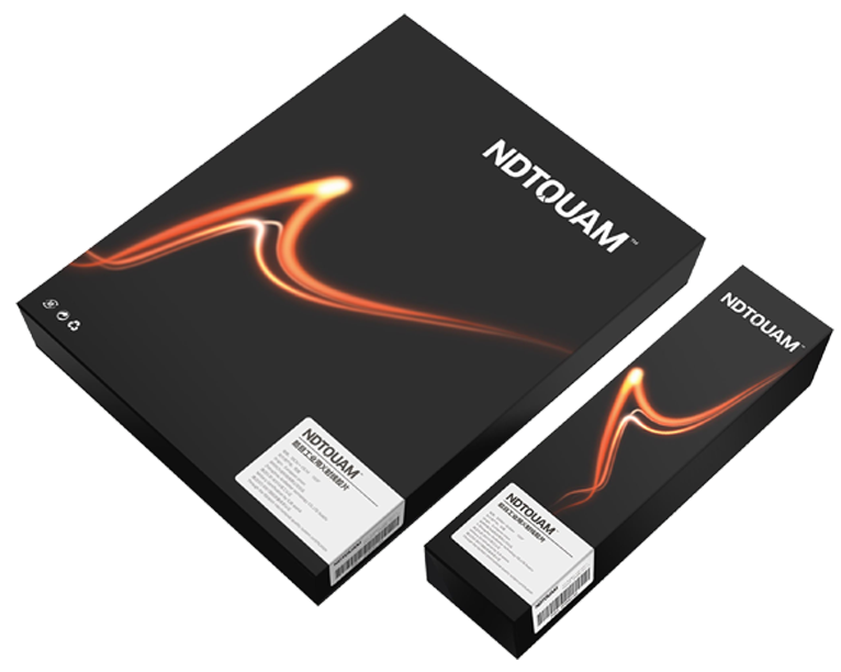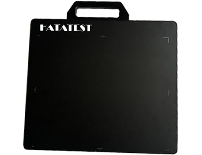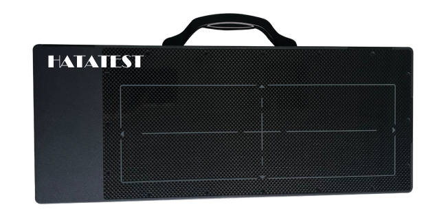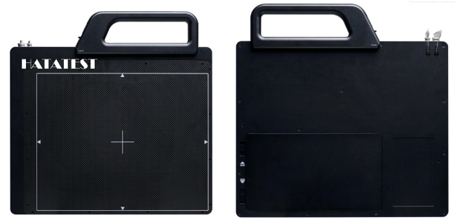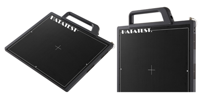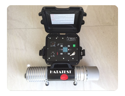
Industrial Digital Radiograp is a classic non-destructive testing technique used primarily to help quality inspectors discover internal defects or discontinuities in some materials. Radiography has been discovered for a long time, and early applications have focused on medical and other fields. However, with the passage of time and the progress of related scientific research, it has been found that this technology has great potential for detection and application in the industrial field (especially in manufacturing).
Today, metal products such as weldments and metal castings are often tested using radiographic technology, which provides manufacturers with great convenience because it not only provides fast, efficient, and accurate product inspection results, but also It does not sacrifice a certain property of the material and damage materials, and is a true non-destructive testing technology.
Nowadays, with the increasing use of polymer materials and the further development of radiographic inspection technology, X-ray inspection technology has become a reliable detection method for many plastic products, silica gel, microchips and rubber.
This test method can be quickly tested to meet various industry specifications. For details, refer to the procedures specified in ASTM (American Society for Testing and Materials), ASME (American Society of Mechanical Engineers), MIL (US Military Standards) and other standards. And specific evaluation criteria. Technicians performing tests and evaluating samples usually need to have a qualification certificate issued by a professional certification body (such as the American Society for Non-Destructive Testing), because only qualified operators will strictly follow the test requirements to ensure qualified test results.

In general, X-ray inspection can be performed in two ways:
(1) Computer digital imaging radiography (DR);
(2) X-ray photography using film imaging (CR) .
CR: An industrial film based on silver bromide (manual or automatic processing) is formed after exposure, and then the captured image is inspected to ensure a sufficiently good lens quality; then the film is evaluated according to the standard.
DR: It is possible to expose an image using a normal X-ray imaging apparatus and to create a digital file. This is primarily achieved by using a phosphor-based imaging plate instead of physical film. The imaging plate is the core component of computer X-ray imaging and is similar to the film action in ordinary X-rays. This method also has the added advantage that the image can be adjusted to increase contrast and to zoom in on specific areas for viewing and more.
Both of the above methods are able to obtain accurate results in a shorter period of time with lower energy; this makes them very accurate and efficient for detecting some special samples.
-
 Sales@hata-ndt.com
 -
 0086-0371-86172891

