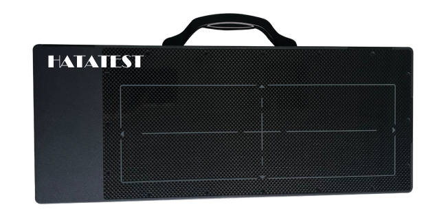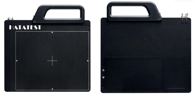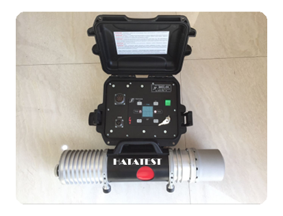
X-ray non-destructive testing has been used in industry for a long time. The early filming method has long time-consuming and high cost, and it has no real-time performance. It is subject to many restrictions in application. In the past ten years, with the development of computer image processing technology, the X-ray real-time imaging system has been rapidly developed. The X-ray real-time imaging system integrates X-ray imaging technology and computer image processing technology into one, and the detection speed is fast. Compared with the traditional filming method, the efficiency of X-ray detection is greatly improved, the detection cost is reduced, and the detection data is easy to save and query, and the real-time dynamic effect is beyond the traditional filmmaking method.
The traditional X-ray real-time imaging system uses an electrostatic focusing X-ray image intensifier, which is bulky and can only be used as a stationary system. The emergence of near-mount X-ray image intensifiers has promoted the development of portable low-intensity X-ray imaging systems, and has emerged as small desktop X-ray images and hand-held X-ray images.
This portable low-intensity X-ray imager offers low power consumption, easy protection, and direct observation in bright environments, providing an important means for clinical inspections, field and field forces in the medical field, as well as in small industrial products. Non-destructive testing, safety inspections in the security sector have also been widely used. However, the imaging quality of this X-ray imaging system is low, and there are few systematic studies to improve its imaging quality. In recent years, the research on its image acquisition and processing technology is limited to the method adopted by large-scale X-ray real-time imaging system. Portable, real-time image processing.
Portable X-ray real-time imaging system consists of X-ray source, single-close image intensifier, CCD, image acquisition and processing equipment, etc. It belongs to tandem imaging system, and the performance of any link in the series system will become the bottleneck restricting system performance.
In order to improve the imaging quality of portable low-intensity X-ray real-time imaging system and expand its application space, this paper systematically studies the key factors affecting the imaging quality of portable X-ray real-time imaging system, and proposes corresponding solutions.
The following research work was carried out in a targeted manner:
(1) The X-ray imaging fuzzy causes and their processing methods are studied. The X-ray geometric imaging performance is evaluated by Tracepro software, which provides a low-cost and efficient solution for the X-ray tube selection in system design. The microchannel plate (MCP) has been theoretically and experimentally studied as an absorption collimator, and the ideal result is obtained. The MCP collimator is small in size and light in weight, and is very suitable for application in a portable X-ray imaging system.
(2) The cathode performance of a single-contact X-ray image intensifier was studied, and the ideal film structure was obtained. The Monte Carlo method was used to simulate the electron scattering on the surface of the cathode. Scattering is an important factor affecting the resolution of near-attach X-ray image intensifiers. A scheme is proposed to reduce the diffusion radius of electrons by using an auxiliary acceleration electric field.
(3) The noise influencing factors of single-contact X-ray image intensifier and CCD are studied. For the real-time imaging system using CCD for image acquisition, the method of reducing the operating voltage of microchannel plate is used to reduce the X-ray image intensifier. Imaging noise, and using refrigeration CCD to reduce the noise during image acquisition, reduce the background noise of CCD by 6 times, and study the resolution matching of X-ray image intensifier and CCD, and give the best pixel matching data.
(4) Aiming at the spatial resolution and contrast sensitivity requirements of portable X-ray real-time imaging system, the image denoising and image enhancement two-step image processing methods are studied. The image denoising processing is performed from the frequency domain, time-frequency domain and spatial filtering angle respectively. Research and comparison show that time-frequency domain filtering has a good effect on X-ray images. By studying various image enhancement algorithms, the contrast stretch transformation using the gray value variation number compression enhancement region can effectively enhance the low contrast X-ray image.
(5) Based on the NIOSII-based FPGA platform, the DSP IP soft core and image processing soft core are designed to realize the parallel structure of image processing, improve the processing speed, and realize the portable real-time image processing function. After optimizing the various parts of the system, the resolution of 6.3 lp/mm was achieved in the X-ray real-time imaging system with single close focus, reaching the level of imported line array and area array.
-
 Sales@hata-ndt.com
 -
 0086-0371-86172891


