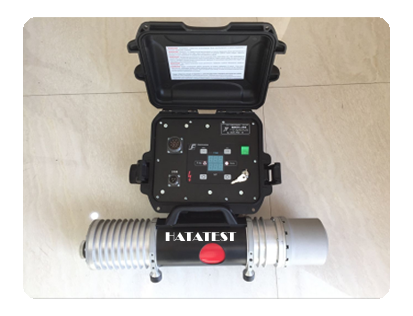
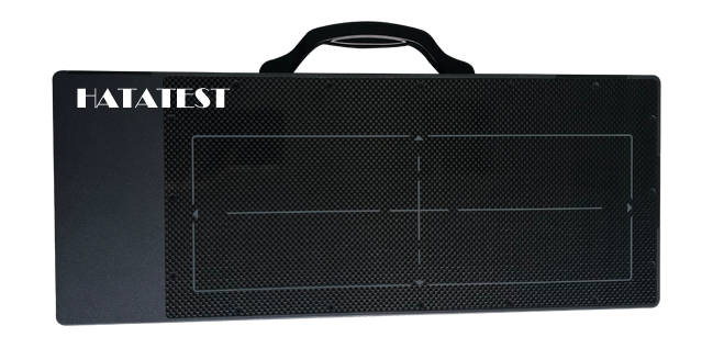
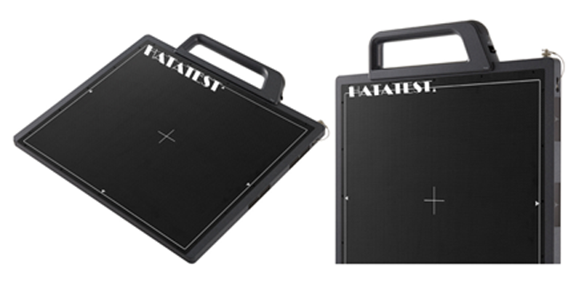
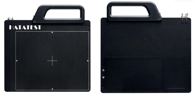
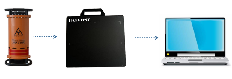
Radiation real-time imaging detection technology refers to the detection technology that can observe the generated image while exposure
1. Development of ray real-time imaging detection technology:
(a) Image brightness enhancer is used instead of a simple screen to achieve image brightness and contrast enhancement.
(b) Radiography is performed by projection magnification using a microfocus or small focus source.
(c) Introducing digital image processing technology to improve image quality.
Implementation of the subordinate conversion process in the image intensifier:
Ray---visible light---electron---visible light

2. Image characteristics of ray real-time imaging system
There are three main indicators of ray real-time imaging system detection system image quality, namely image resolution, image unsharpness and contrast sensitivity. These three indicators roughly correspond to the graininess, unsharpness and contrast of film photography, but the imaging principles, methods and equipment of real-time imaging and film photography are different.
(a) Image resolution. Image resolution, also known as spatial resolution, is defined as the minimum spacing of the display screen image to identify line separation, in Lp/mm (pair/mm). The image resolution of the ray real-time imaging inspection system can be measured by a line-to-test test card, and the platinum-tungsten dual-wire image quality meter can also be used to determine the image resolution.
(b) The image is not clear. After imaging a device with a sharp boundary, it affects the width of the blurred area of the boundary. The factors that affect the image's unsharpness are mainly the geometric unsharpness and the inherent unsharpness of the screen. In ray real-time imaging detection techniques, geometric unsharpness is related to the selected magnification, in addition to the focus size. The image unsharpness can be measured by a line-to-test test card or by a two-wire image quality meter. The image of the ray real-time imaging detection system is easy to achieve high contrast, but often does not get satisfactory clarity.
(c) Comparison sensitivity. Contrast sensitivity is defined as the percentage of transillumination thickness identifiable from the display image, ie ΔT/T. The contrast C of the image observed on the display of the ray real-time imaging inspection system is related to the main cause contrast and the brightness of the screen. The contrast sensitivity can be determined using a step test block.
3. Process points of ray real-time imaging detection technology
(a) Best magnification
The image is enlarged to facilitate the identification of small defects. However, as the magnification increases, the geometric unsharpness also increases, which is not conducive to defect recognition.
(b) Scanning speed and positioning accuracy
The ray real-time imaging inspection process includes dynamic inspection and static inspection. For dynamic testing, it is necessary to control the moving speed of the workpiece and the ray source, which is directly related to the noise of the image.
(c) Image processing. Digital image processing techniques used in ray real-time imaging detection techniques include contrast enhancement (gray enhancement), image smoothing (multi-frame averaging noise reduction), image sharpening (boundary sharpening), and pseudo-color display.
(d) System performance verification. In order to ensure reliable test results, the performance of the system must be periodically verified. There are two types of verification methods: static check and dynamic check. Static calibration items include image resolution and contrast sensitivity. The period and interval of calibration should meet the relevant requirements. When using the defective sample for dynamic verification, the transillumination parameters and the moving speed of the test piece should be the same as the actual test. The selection, number and placement of the image quality meter should conform to the standards and process specifications.
-
 Sales@hata-ndt.com
 -
 +86 371 63217179

